Spontaneously hypertensive rats provide a model of genetic hypertension that allows the study of primary hypertension. The BIBW2992 EGFR/HER2 inhibitor administration of carbohydrate-rich diets to rats can induce insulin resistance, hyperinsulinemia, dyslipidemia and moderate hypertension. Chronic fructose-fed rats provide a useful GANT61 experimental model for studying the interaction of the factors that shape metabolic syndrome. We postulate that this dual experimental model could be appropriate for extrapolating results to human pathology. In the present study, 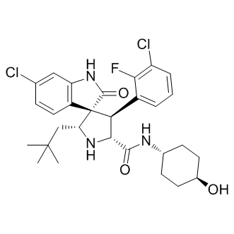 we used vidagliptin to examine the role of the DDP-IV, incretin system component, in the activation of different molecular inflammatory cytokines, NF-kB and VCAM-1 to generate a microenvironment that supports cardiovascular remodeling. In this article, we demonstrated an important effect of DDP-IV in reducing vascular inflammation, accompanied by a favorable reduction in metabolic and structural parameters. The FFHR experimental model presents hypertension, dyslipidemia, insulin resistance, vascular and cardiac remodeling, inflammation demonstrated by increased hsCRP and vascular inflammation due to increased expression of NF-kB, VCAM-1 and pro-atherogenic cytokines. The increased expression of VCAM-1 is a marker of vascular inflammation, vascular permeability and endothelial dysfunction. The inflammatory process identified in this experimental model has two components: 1- a local component involving an increase in the levels of nuclear transcription factors with subsequent activation of the inflammatory cascade, resulting in a strong presence of cytokines, and level 2�C a systemic component involving increased hepatic synthesis of CRP due to a probable increase of IL-6. The data suggest that incretin system dysfunction, as happen in patients with diabetes mellitus or metabolic syndrome, allows activation of inflammatory response in different levels. With the consequent creation of a vascular microenvironment that is conducive to the creation, perpetuation, progression, and destabilization of vascular injury, with either a simple eutrophic mechanism of vascular remodeling, or the generation of an atherosclerotic lesion. A number of mechanisms may underlie these results. Given that GLP-1 is a physiological substrate of DPP-IV, DPP-IV inhibition by V may be expected to increase the circulating levels of GLP-1 Several studies have reported beneficial effects of GLP-1 on the cardiovascular system. In humans, Nikolaidis et al. have shown that a 72-h infusion of GLP-1 improved left ventricular function in patients with acute myocardial infarction and systolic dysfunction after successful reperfusion therapy, an effect that was observed in both diabetic and nondiabetic patients. The authors suggested that this observation might be explained by the insulinotropic and insulinomimetic properties of GLP-1; alternatively, GLP-1 might also improve endothelial function. Studies have shown that GLP-1 improves endothelium-dependent vascular responses in the brachial artery while leaving endotheliumindependent responses unaffected in healthy humans and patients with type 2 diabetes. The cardiovascular actions of GLP-1 may occur either directly through the GLP-1 receptor or through a GLP-1 receptor-independent effect of the degradation product of GLP-1, GLP-1. In addition to GLP-1, DPP-IV also degrades GIP, and potentially cytokines and certain chemokines. Thus, other substrates of DPP-IV may be responsible for the improvement in endothelial function. Alternatively, V might improve endothelial function by influencing insulin and glucose levels. Insulin causes vasodilatation by increasing endothelial production of NO.
Each biological replicate within an experiment is indicated allowing for variability of each peptide among replicates to be compared
The heat map analysis shown in XL-184 Figure 5 excludes AM114, which did not substantially inhibit the proteasome at the concentration used in the peptidomics study. Peptides selected for inclusion in the heat map were chosen based on the number of times found in each of the distinct experiments; peptides found in at least five separate runs are included. Only a handful of peptides were detected in every replicate in every experiment. The failure to detect a peptide doesn’t necessarily mean it isn’t present; there are multiple reasons for the absence of a signal. In general, the studies with MG132, MG262, and clasto-Lactacystin b-lactone resulted in fewer detectable peptides than the other studies. Despite this limitation, several trends were detected in the heat map analysis. First, many of the same peptides elevated upon treatment of cells with bortezomib are also elevated by MG262. In contrast, most of the other proteasome inhibitors do not cause these peptides to be elevated. One exception is carfilzomib, which produces an increase in some but not all of the peptides elevated by bortezomib and MG262. Another trend revealed by the heat map analysis is that some peptides show similar responses to all of the proteasome inhibitors. One set of peptides was decreased in at least five of the runs and had an average ratio in all runs of #0.65. In some replicates, these peptides were in the “no change” group, but never showed an increase in any of the replicates. Altogether there were 18 peptides in this set. The majority of these peptides represent the N-terminus or C-terminus of the protein, and therefore only a single cleavage is required to produce the peptide. All of the peptides in this set are produced by cleavages attributed to the beta 5 proteasome subunit, based on the presence of a hydrophobic amino acid residue in the P1 positions of the cleavage sites required to generate the peptide. Another set of peptides was not greatly affected by the proteasome inhibitors in any of the replicates. The average ratio for these peptides ranged from 0.85 to 0.99 in all of the studies, and in no case was a large change found in any of the replicates. One member of this group is the small protein thymosin beta-10, which only undergoes removal of the initiation methionine and would not be expected to be altered by treatment with proteasome inhibitors. A third set of peptides was found to increase in at least five of the experiments, with an average ratio.3.0 for all experiments, and no replicate WY 14643 showing a decrease in any of the experiments. Of the 11 peptides in this group, the majority represented internal fragments of the protein and therefore required two cleavages to be produced. Of the 18 cleavage sites required to produce these peptides, only 11 match the consensus site for beta 5 cleavages, the rest match the consensus site for beta 1 or beta 2 cleavages. There was no substantial difference in the average mass or peptide length for the peptides in set 1 versus set 3. The finding that bortezomib and other compounds increase the levels of some peptides can be explained by one of two possible mechanisms; either the compounds increase the formation of the peptides or the compounds block the degradation of the peptides. A recent study predicted that bortezomib could inhibit TPP2. TPP2 is thought to play a major role in peptide degradation within the cell. To test whether bortezomib inhibited TPP2, we first assayed HEK293T cell extracts with the TPP2 substrate AlaAla-Phe-AMC. Because this substrate is not specific for TPP2 and can be degraded by other cellular peptidases, we examined the activity in the presence of various concentrations of the TPP2-selective  inhibitor butabindide.
The development of a high-resolution genetic linkage map is many types of biotic and abiotic stresses
The outermost wax layer protects plants from such as drought, phytophagous insects, pathogens, solar radiation, and freezing temperatures. One of the most important roles of the cuticle is  to limit transpiration to reduce water loss and this provides a key mechanism for plant survival in water-limited environments, such as deserts, high mountains, saline-alkali lands, and coastal ecosystems. Worldwide, bread wheat is one of the most important food sources for human beings. The wheat leaf, stem and, in some cases, spike surfaces are coated with cuticular waxes that confer a glaucousness characteristic. Physiological studies in wheat by Johnson et al. and Richards et al. showed that glaucousness reduces transpiration and increased water use efficiency. More recently Zhang et al. demonstrated that glaucousness reduced cuticle permeability in the terms of nonstomatal water loss and chlorophyll efflux. Bread wheat cultivars with non-glaucousness traits exhibit significant yield increases with reduced solar radiation losses that enable continued photosynthesis during the grain filling period, and the trait may also provide resistance to aphids. Glaucousness and non-glaucousness are parallel variations in wheat and its relatives. Classical SAR131675 molecular weight Genetic studies have shown that both the glaucousness and the non-glaucousness stem and leaf phenotypes are controlled by two sets of loci; the wax production genes W1 and W2 and the wax inhibitor genes Iw1 and Iw2, respectively. The Iw1 and Iw2 non-glaucousness loci function as inhibitors of the W1 and W2 glaucousness loci, and could also inhibit other wax production genes in the wax pathway. Genetic analyses have indicated that the W1 wax production gene and the Iw1 wax inhibition gene are located on chromosome 2BS with a genetic distance of 2 cM. However, W2 and Iw2 are separated on chromosome 2DS where the W2 locus is close to the centromere. Two loci, Iw3 and Ws, were also reported conditioning wax on spikes in wheat. Non-glaucousness locus Iw3 was mapped on chromosome 1BS and the Ws gene on the short arm of chromosome 1AS is responsible for glaucous spikes. In addition to these genes, a major QTL that accounts for up to 52 percent of the flag leaf glaucousness variation has been detected in a doubled-haploid population. Molecular mapping and cloning of genes controlling epicuticular wax in wheat is of great interests for understanding interactions between none-glaucousness genes and glaucousness genes, as well as their effects on yield, and biotic and abiotic stresses. The Iw1 locus originating in wild emmer is closely linked to the Xcdo456 RFLP marker at the end of chromosome arm 2BS. Liu et al. found that the Iw1 locus is 18.77 cM away from the powdery mildew resistance gene MlIW170 on chromosome 2BS. Simmonds et al. also reported that the Iw1 gene conditioning a non-glaucousness phenotype maps to chromosome 2BS. In a tetraploid wheat background, Yoshiya et al., have found that W1 is linked to Iw1Dic, but the relationship between Iw1 and Iw1Dic was not confirmed, and in an Ae. tauschii F2 segregating population, the non-glaucous locus Iw2 was located on chromosome 2DS. In another report, the dominant non-glaucous locus Iw3672 derived from a AZ 960 synthetic hexaploid wheat also mapped on 2DS by simple sequence repeat and expressed sequence tag markers. During development of a wheat genetic linkage map with a doubled haploid population derived from the TA4152�C60 synthetic hexaploid wheat line and the ND495 common wheat line, Chu et al., also located a dominant wax inhibitor Iw2 on chromosome 2DS. Compared to studies on the Iw nonglaucousness loci, little work has been done to map the W glaucousness loci in wheat, aside from W1, which has been mapped on chromosome 2BS and the Ws glaucous spike allele that is located at the terminus of chromosome 1AS.
Endothelial progenitor cell proliferation and differentiation towards the endothelial cell lineage in vitro
Our previous study showed that high glucose levels did not alter apoptosis in endothelial progenitor cells in vitro, but activated p38 MAP kinase and several downstream targets such as the transcription factor CREB. Unfortunately, no animal studies in models for type 2 diabetes or metabolic syndrome were conducted to identify signalling mechanisms in vivo which contribute to high glucose-induced impairment of progenitor cells. One critical issue is to choose an appropriate mouse model that closely resembles type 2 diabetes in humans, which is the major form of diabetes and accounts for 95% of diabetes cases. There are very few animal studies to identify signalling mechanisms of endothelial progenitor cells in diabetes. It was shown that NO-mediated impaired mobilization may be responsible for the progenitor cell reduction in diabetic mice. However, Gallagher et al. used streptozocin-treated mice, a model not comparable with type 2 diabetes in humans. To study type 2 diabetes in a mouse model we chose the leptin receptor knock out mouse model. Leprdb mice lack functional leptin receptors, become obese shortly after birth and are insulin resistant and hyperglycemic as adults. The aim of this study was to investigate potential signalling mechanisms in vasculogenic progenitor cells,Avitinib maleate which regulate the differentiation processes in vitro and in vivo. ETS is one of the largest families of transcriptional regulators that share a highly conserved DNA-binding domain. ETS transcription factors are downstream of the p38 MAP kinase, which has already been shown to be important for endothelial progenitor cell differentiation and proliferation. As ETS transcription factors are important for proliferation, survival and differentiation, we hypothesized that this could also be an important mechanism in VPC. We found that ETS DNA-binding activity is much higher in diabetic cells. Inhibition of the ETS activity leads to increase in impaired VPC number. Therefore this could be a potential mechanism to improve the impaired VPC number in patients with type 2 diabetes. The data of the present study demonstrate that increased nuclear DNA-binding activities of ETS1 and ETS2 transcription factors are crucial determinants of diabetes- and high glucoseinduced reduced number of VPC. Moreover,DprE1-IN-1 increased DNAbinding activity of ETS-transcription factors inhibits the commitment of progenitor cells towards the endothelial cell lineage. Emerging evidence indicates that endothelial progenitor cells are involved in the maintenance of vascular homeostasis and their impairment may be conducive to vascular disease. Endothelial progenitor cell function is impaired in patients with coronary artery disease and diabetes. Recently, Fadini et al. suggested that diabetes is the most relevant risk factor associated with a reduction of endothelial progenitor cells. However, the underlying mechanisms are still not understood. Marchetti et al investigated the effects of high glucose toxicity on endothelial progenitor cells and determined that high glucose reduced phosphorylation of Akt and thereby increased activation and nuclear localization of the forkhead transcription factor-1 previously shown to have negative impact on endothelial function.
The isoforms HDAC1 and HDAC4 were demonstrated as critical in regulating acetylated HMGB1 release
The hepatocytes with nuclear immunoreaction for acetylated-lysine were increased in number 1–3 h after the concomitant administration of LPS/GalN, but they were decreased. At 8 h, aberrant, extracellular acetylated-lysine expression was labeled and mainly restricted to pericentral inflammatory areas. At 10 h a distinct nuclear expression for acetylated-lysine was again increased in the hepatic cells, although some hepatocytes remained to express cytoplasmic acetylatedlysine in the pericentral areas. Administration of GL strongly inhibited the immunoreaction of acetylated-lysine in the LPS/GalN-induced liver injury. Immunohistochemical analysis for acetylated-lysine used in this experiment shows that administration of GL may Fruquintinib inhibit acetylation of lysine in HMGB1 in the liver remnants of the LPS/GalN-induced mice. The ability of GL to suppress ALT levels is observed when administered at 30nmin before or at 10 min and 60 min after LPS/GalN, but GLtreatment has little effect on ALT levels when administered 3 h after LPS/GalN injection. Thus, inhibitory effect of GL on LPS/GalN-induced liver injury might be due to binding directly to HMGB1 before its acetylation. Acetylated HMGB1 has been shown to be involved in regulating HMGB1 DNA binding properties along with its subcellular location. In vitro experiments have shown that lysine residues of HMGB1 between 27 and 43 represent functional nuclear localizing signals in macrophages. Also, recent mutation analysis for the abilities of HMGB1 protein to bind and bend DNA has reported the role of lysines 2 and 81 as target sites for acetylation in full-length HMGB1 and truncated tail-less protein,Farampator respectively. Serum HMGB1 released following liver ischemia/reperfusion in vivo is acetylated and hepatocytes exposed to oxidative stress in vitro also release acetylated HMGB1. Levels of acetylated HMGB1 increase with a concomitant decrease in total nuclear histone deacetylase activity, suggesting that suppression in HDAC activity contributes to the increase in acetylated HMGB1 release after oxidative stress in hepatocytes. Activation of HDAC1 is decreased in the nucleus of hepatocytes undergoing oxidative stress. In addition, HDAC1 knockdown with siRNA promotes HMGB1 translocation and release. Furthermore, HDAC4 is shuttled from the nucleus to cytoplasm in response to oxidative stress, resulting in decreased HDAC activity in the nucleus. On the contrary, in the present experiment we did not find a decrease in both the cytoplasmic and nuclear HDAC activities after injection of LPS/GalN. In addition, administration of GL did not induce a significant increase of nuclear HDAC activity. Phosphorylation of HMGB1 is another regulatory mechanism that influences its subcellular location. Recently, it was shown that HMGB1 phosphorylation by calcium/calmodulin protein kinase IV caused nuclear-to-cytoplasmic shuttling and release in LPS-stimulated macrophages. Damaged cells activate innate immunity and recruit inflammatory cells by collectively releasing danger signals known as the damage-associated molecular patterns. One such molecule is HMGB1, which has been implicated as an early mediator of organ damage in ischemia/reperfusion injury and hemorrhagic shock, as well as a late mediator of lethality in endotoxic shock. In most cases the cells actively secreting HMGB1 appear to be immune cells such as macrophages, natural killer cells, and dendritic cells. However, it is becoming increasingly clear that non-immune parenchymal cells also participate in active HMGB1 secretion.