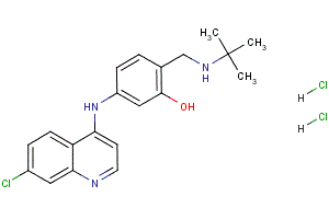
Interestingly, ER stress signaling has been shown to be a negative regulator of malignancy in human squamous carcinoma cells and of H-Ras-mediated transformation of human melanocytes. Further, inhibition of PKR and subsequent reduced 20S-Notoginsenoside-R2 phosphorylation of eIF2a was sufficient to cause transformation of mouse NIH3T3 fibroblasts. These results suggest that phosphorylation of eIF2a could potentially have a tumor inhibitory function. Our results reveal two important and previously unrecognized salient findings: the first is a link between adhesion signaling and PERK-dependent phosphorylation of eIF2a; the second, is the adhesion-dependent role of PERK in inhibiting proliferation and tumor formation both in 3D in vitro and in vivo animal models, respectively. Early studies showed that MEFs in suspension displayed translational repression. More recent studies showed that in NIH3T3 fibroblasts, the suspension-induced translation repression correlated with increased P-eIF2a levels. Our results are in accordance with, and further extend these findings by showing that loss of adhesion can regulate eIF2a phosphorylation in a PERK-dependent manner. That PERK is responsible for eIF2a phosphorylation, is supported by our results showing that PERK-/MEFs display reduced phosphorylation of eIF2a as compared to wtPERK MEFs placed in suspension. Importantly, activation or inhibition of PERK-independent pathways appeared to strongly regulate suspension-induced translational repression, as inhibition of PERK was not sufficient to restore protein synthesis and prevent anoikis. Alternatively, this could be due to residual eIF2a phosphorylation, since as little as 10% of eIF2a phosphorylation can cause a strong repression of translation. Another possibility maybe the fact that the eIF4E pathway may still be inhibited in suspension. It is also possible that phosphorylation of PERK and eIF2a in suspension may have other functions not immediately linked to apoptosis but to other forms of cell death. Moreover, it was interesting to find that PERKDC enhanced protein synthesis in adhered conditions and prevented DTT-induced translational repression. This suggests that PERK signaling may be required in the adherent cells to inhibit proliferation and PERKDC-expressing cells might be refractory to these signals during acinus formation. Perhaps, maintenance of LN-5 expression at basal levels is required to prevent hyperproliferation as only PERKDC MCF10A cells were surrounded by large LN-5 deposits and positive for Ki67 in the lumen-filled acini. The signals that activate PERK during acinar morphogenesis are unknown but it is possible that more subtle changes in adhesion or in matrix composition that activate PERK may become evident as the acinus reaches its terminal size. Our results showed that the proliferative and tumor suppressive effect of PERK and eIF2a signaling is not due to their ability to induce anoikis in response to loss of adhesion, but due to the inhibition of proliferation in adherent cells. The PERKDCinduced stimulation of proliferation and tumor initiation was Alprostadil unexpected and raises the question of the mechanisms behind this phenomenon. The PERKDC-induced phenotype resembled, although not as strongly, that of ErbB2 activation in MCF10A cells. To conclude, our studies reveal that PERK activation and possibly downstream eIF2a signaling is regulated by adhesion through an as yet unknown mechanism. This signal appears to be required to limit proliferation and allow for normal acinar  morphogenesis. Most importantly, this pathway appears to have a tumor suppressive effect.